

dr Anton Friedman

Witten, Germany

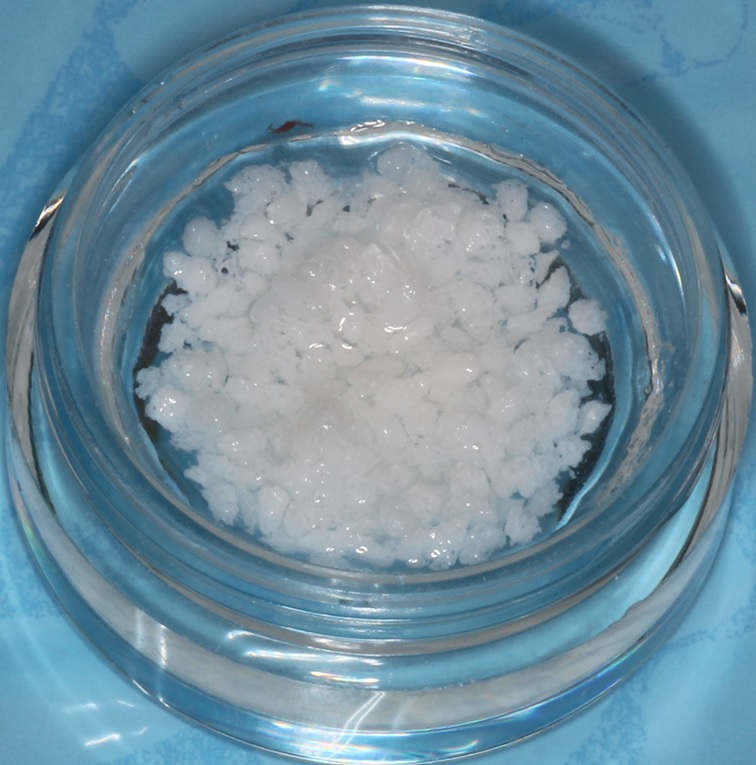
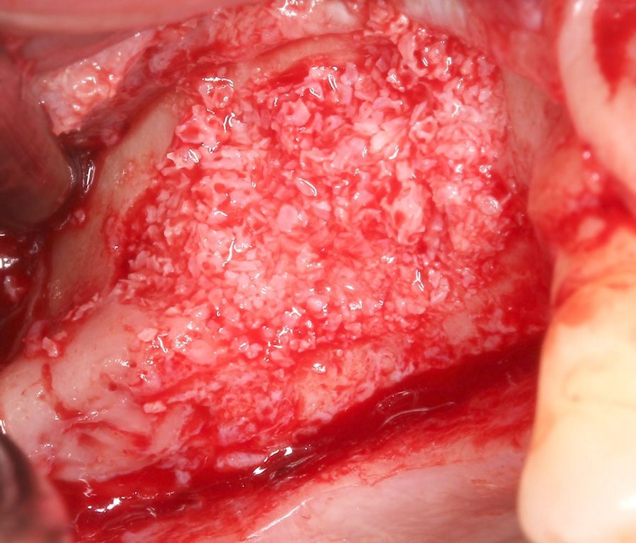
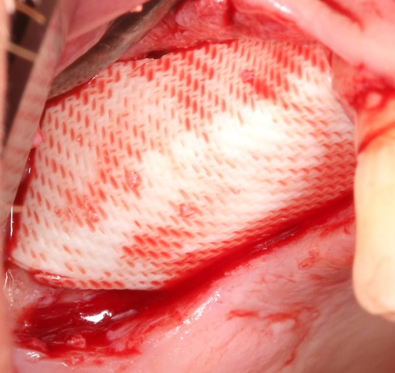
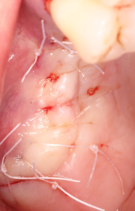
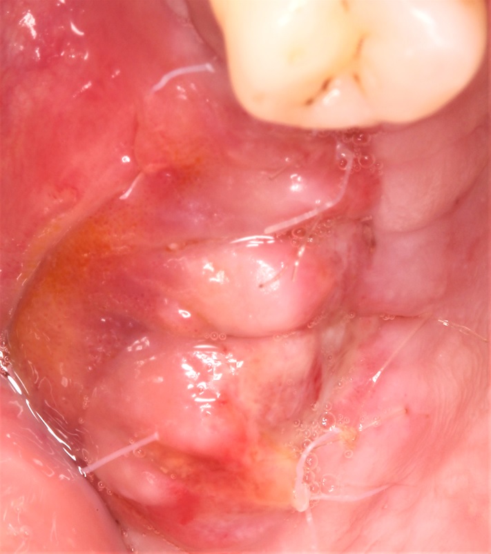
SmartGraft
 Zobacz produkty użyte
 w tym przykładzie


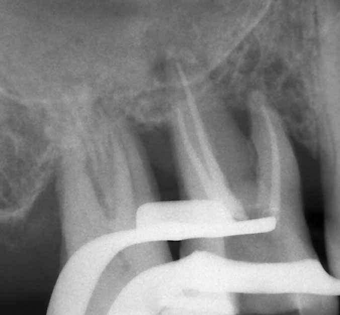
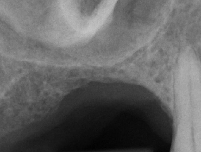
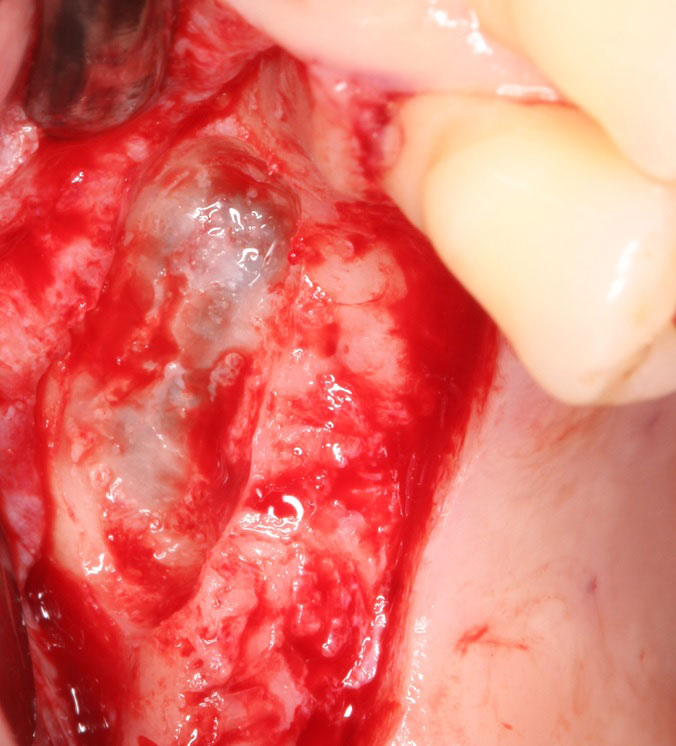
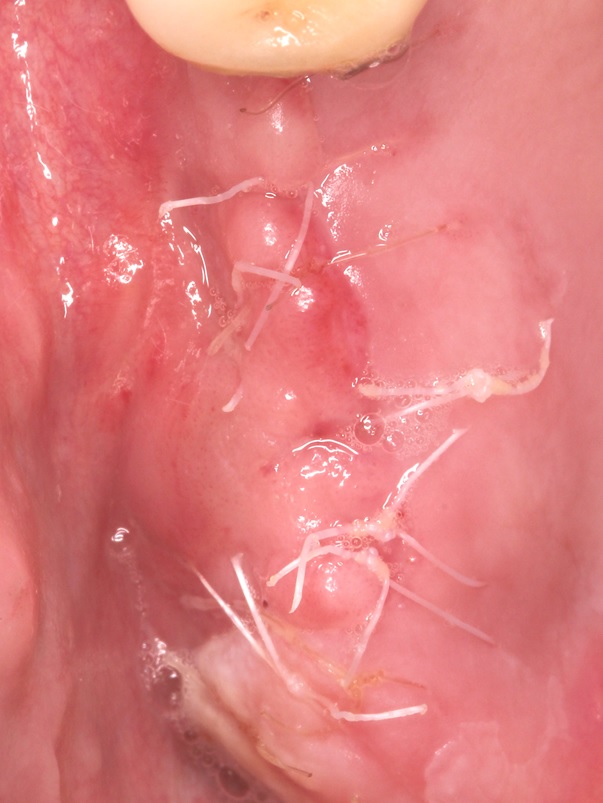
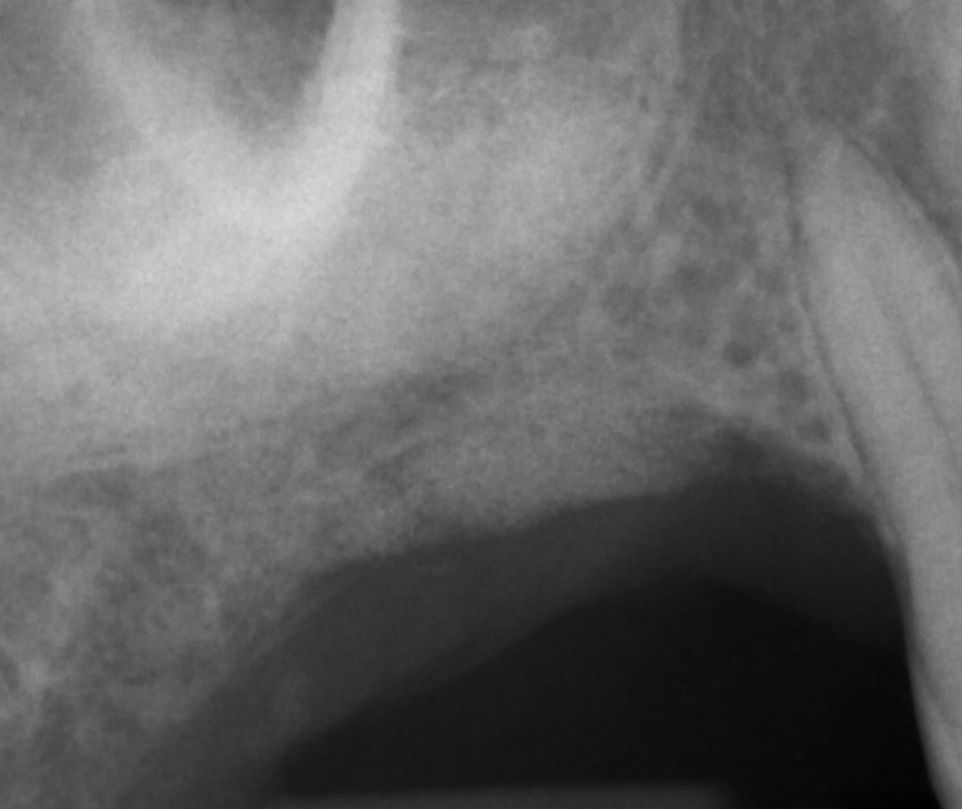
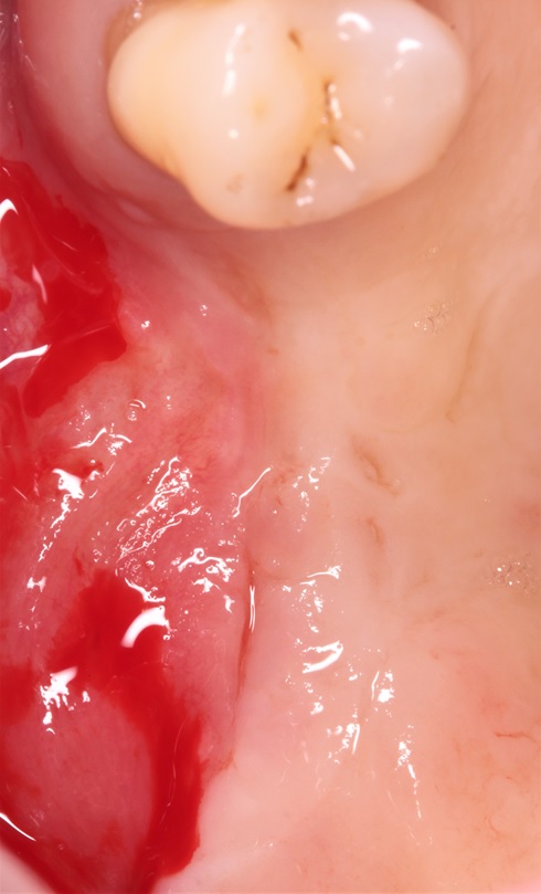
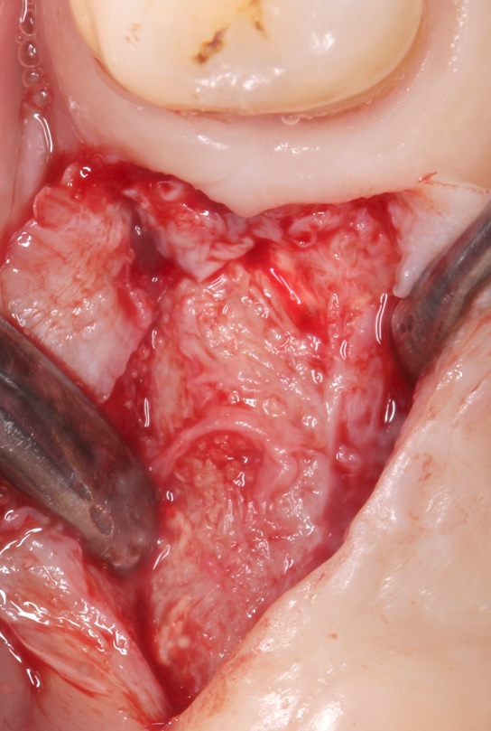
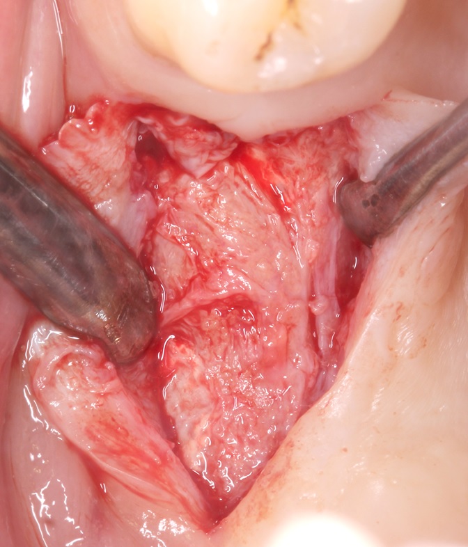
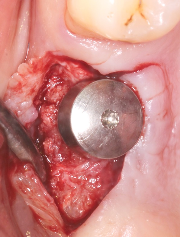
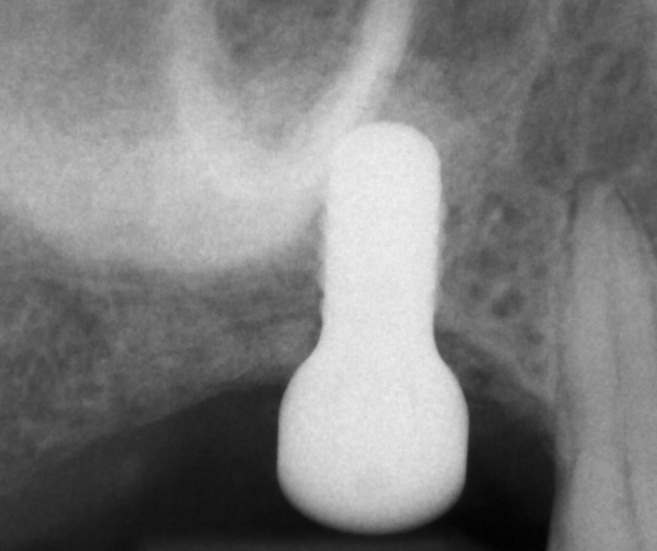
Case provided by prof. dr. Anton Friedman (Chair and Head Department of Periodontology, School of Dentistry, Faculty of Health, University of Witten, Germany).
dr Anton Friedman
Witten, Germany
SmartGraft
Zobacz produkty użyte WSPÓŁPRACA:

Zgodnie z obowiązującymi przepisami prawa, niniejsza strona i prezentowane na niej treści są przeznaczone wyłącznie dla profesjonalistów tzn. w szczególności osób wykonujących zawód medyczny lub w inny sposób związanych zawodowo z branżą medyczną.
Aby przejść dalej, prosimy o potwierdzenie czy jesteś profesjonalistą.